
Macular Degeneration

Macular Degeneration affects approximately 25% of individuals over 50 years of age. This degenerative condition is caused by inflammation to the tissues and nerves in the eye. This results in deterioration in the central retina (macula) and affects the central vision, leaving peripheral vision intact.
The macula is part of the retina located in the back of the eye and is responsible for central vision. It is the only part of the retina capable of seeing fine detail. As a result, normal daily activities such as reading and driving are affected by this disease. Total blindness rarely occurs from macular degeneration. Although the exact cause of macular degeneration is not known, risk factors include advancing age, family history, exposure to UV light, and smoking. Nutritional factors such as deficiency in vitamin B, E, zinc, and/or Magnesium intake may also play a role.
Macular Degeneration Symptoms:
- Straight lines appear wavy, crooked or disconnected
- Dark or blank areas appear in the center of your vision
An Amsler Grid can be used to help monitor your vision for macular degeneration. While wearing the glasses you would normally wear, cover one eye and look at the black spot in the center of the square. If you see any distortion or dark areas in the square you need to call your eye physician immediately.

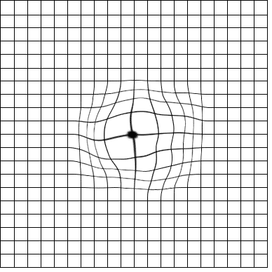
The two forms of macular degeneration are dry and wet, with 90% of cases being dry.
Dry Macular Degeneration
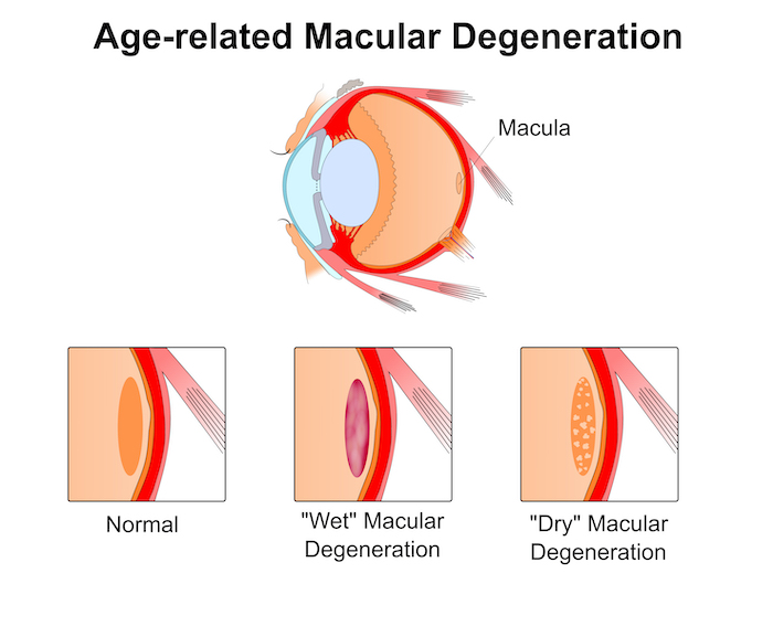
Dry macular degeneration is characterized by a slow deterioration in vision. Drusen are the clinical hallmark of the dry form of macular degeneration. These are metabolic by-products (accumulated retinal waste) that are deposited in the central retina, causing the retina to become thinner. This type of Macular Degeneration causes a slow deterioration in the light sensitive cells in the macula. Because there is no leakage, this form is considered “dry”. Patients with dry macular degeneration should be monitored very closely for conversion to the wet form.
Wet Macular Degeneration
The wet form of macular degeneration tends to be more visually aggressive. Wet macular degeneration occurs when new blood vessels grow beneath the oxygen-deprived retina. These new vessels are very fragile and leaky, bringing damage to the surrounding tissue and causing the retina to lift up. When this happens the central vision becomes distorted. This type of Macular Degeneration may cause a sudden loss of vision Or a slow progression of symptoms over time.
Treatment for wet macular degeneration is aimed towards sealing the leaking or bleeding blood vessels. These treatments include Lucentis, Avastin, Macugen, and laser.
Recommendations for Macular Degeneration patients:
- Monitor your vision weekly with an amsler grid. This grid aids in early detection of changes that may require treatment. Report any changes in its appearance to your doctor immediately.
- Take a good nutritional supplement such as Lutein that includes antioxidants and Omega-3 fatty acids.
- Wear sunglasses with an ultraviolet (U.V.) filter to protect the eyes from the damaging rays of the sun.
- Do not smoke.
Diabetic Retinopathy
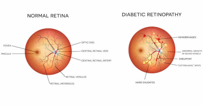
As a diabetic patient, it is important to be monitored yearly for changes in the retina. Prolonged high blood sugar levels may result in damage to blood vessels in the retina causing them to leak.
Diabetic retinopathy progresses from the early stage of the disease, known as non-proliferative, to the later stage, known as proliferative. In the non-proliferative stage, the blood vessels begin to leak, causing the retina to swell and produce blurry vision. If the disease progresses to the proliferative stage, both central and peripheral vision are affected. The blood vessels in the retina become weaker, carrying less oxygen and nutrients to the retinal tissue. In turn, the retina begins to grow abnormal new blood vessels which are fragile and hemorrhage easily. If left untreated, the bleeding will produce scarring and possible retinal detachment.
Diabetic Retinopathy Symptoms
Because diabetic retinopathy often develops with few or no symptoms, many patients go undiagnosed and find themselves left with no treatment options by the time they discover the disease. Early detection is critical, which is why yearly dilated exams are recommended to all diabetic patients.
Floaters
What are Floaters?
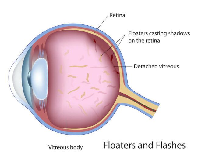
Floaters are seen as spots, specks or flecks that drift into the field of vision. This is very common, especially as we age, and typically are not a cause for immediate concern unless excessive.
The space located between the lens of the eye and the retina is filled with a material called vitreous, which is similar to clear jello. As we age, the normal jello-like consistency of the vitreous begins to liquefy and pull away from its natural attachments on the inside surface of the eye. This causes gel particles to become loose and float around the eye. It may appear that these specks are on the front surface of your eye, but they are actually inside.
Are Floaters Dangerous?
Detachment of vitreous is not by itself dangerous, Except in rare circumstances, floaters are no cause for alarm and no treatment is necessary. However, it can be accompanied by more serious eye conditions such as retinal tears and vitreous hemorrhage. These occur when the strong attachments of the vitreous to the retina do not separate properly, tearing the retina or retinal blood vessels. This often leads to new floaters and persistent light flashes.
We suggest that anyone with symptoms of a vitreous detachment have an eye examination to make certain that a more serious problem is not present.
If you would like to book your appointment online click here or call (561) 338-7722 today!
